Figure 5.
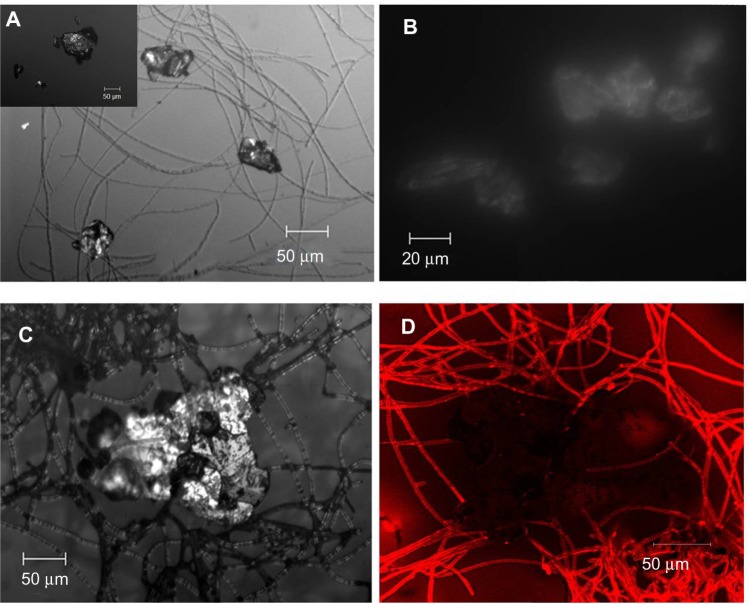
Confocal micrographs of GC7 showing G clusters attached to CL strands (A) (GW shown in the inset), self-assembly of G flakes (B), CL attack at the G edges (C), and fluorescent image showing presence of CL within G (D).
Notes: The corresponding fluorescence image in Figure 5D shows that the large G flake (~200 μm) is not only surrounded by the fluorescing CL fibrils, but the fibrils have even penetrated the flakes from all sides.
Abbreviations: CL, collagen; G, graphene; GC7, 7-day graphite/collagen dispersion; GW, graphite dispersed in water.

