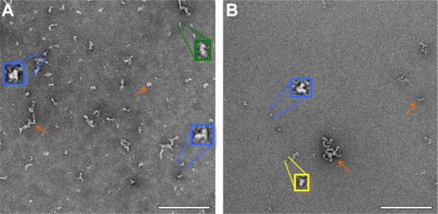
Figure 2.

Negative stain TEM of ID93. Negative stain TEM images of ID93 in 20 mM Tris buffer at different protein concentrations indicate the presence of various structures, including small linear particles (yellow), medium linear particles (green), tripod-shaped particles (blue), and particle clusters (orange arrows). The samples were imaged at 52,000× magnification with insets shown at larger scale. (A) 0.5 mg/mL ID93. (B) 0.05 mg/mL ID93. Scale bar =200 nm.
Abbreviation: TEM, transmission electron microscopy.
