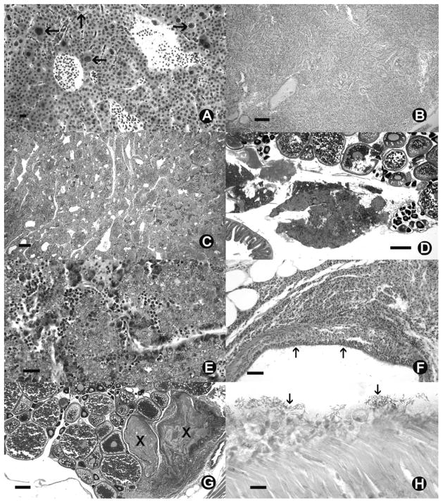
Fig 1.
Danio rerio Histological sections of zebrafish from SPF colony. (A) Severe hepatocytic megalocytosis, with enlarged nuclei (arrows) and hepatocytes. (B) Cholangiocellular carcinoma replacing entire liver parenchyma. (C) Spermacytic seminoma. (D) Ovotestes. S = seminoma in mesenteries adjacent to mature ovaries. (E) High magnification of seminoma from fish with ovotestes. (F) Chronic aerocystitis. (G) Oophoritis (egg associated inflammation and fibroplasia) X = raft of eosinophilic yolk debris and fibroplasia. (H) Swimmbladder surface with acid bacteria (arrows). (A, H) = 10 μm. (B, C) = 100 μm. (F) = 50 μm. (E) = 25 μm (D, G) = 500 μm. (A–E) = Hematoxylin and eosin. (H) = Fite’s acid fast.

