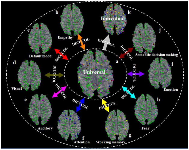
Fig. 1.
Overview of the DICCCOL brain reference and localization system (Zhu et al., 2012). Spheres in orange (total 6), red (total 8), brown (total 9), pink (total 8), blue (total 27), yellow (total 14), cyan (total 14), purple (total 16), and black-red (total 19) colors represent DICCCOLs in empathy, default mode, visual, auditory, attention, working memory, fear, emotion, and semantic decision making networks that are identified from fMRI datasets (Zhu et al., 2012). The green spheres (totally 263) are landmarks that have not been functionally-labeled yet. The DICCCOLs can be used as the structural substrates to represent the common structural brain architecture. For instance, nine functionally-specialized networks ((b)–(j)) identified from different fMRI datasets (Zhu et al., 2012) can be integrated into the same universal brain reference system (a) via DICCCOL. Also, the functionally-annotated DICCCOLs can be predicted in an individual brain with DTI data such that the DICCCOLs and their functional roles can be readily transferred to a separate brain image (k).

