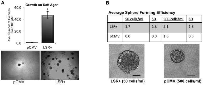
Figure 6. LSR enhances cell survival in non-adherent culture conditions.
Hs578t cells were stably transfected with either a control plasmid (pCMV), or a plasmid containing the full-length gene for LSR variant 1 (LSR+). (A) Soft agar assays. Cells were plated on soft agar coated wells, grown for seven days, then stained with nitrobluetetrazolium before counting. The entire dish was analyzed and colonies larger than 50 um in diameter were counted. Data represent mean colonies counted per well ± SE of three separate experiments; *P<0.001. Bottom panels are representative images at 10X. (B) Sphere forming efficiency. Cells were plated in DMEM +10 ng/ml EGF +20 ng/ml FGF +2% B27 in ultralow attachment dishes for seven days then spheres counted and imaged. Data represent mean +/− SD of three independent experiments at the indicated cell plating densities. Bottom panels are representative phase images of anchorage-independent, single-cell derived spheres from LSR+ and control pCMV cells after seven days of growth. Scale bar, 50 um.

