
Fig. 4.
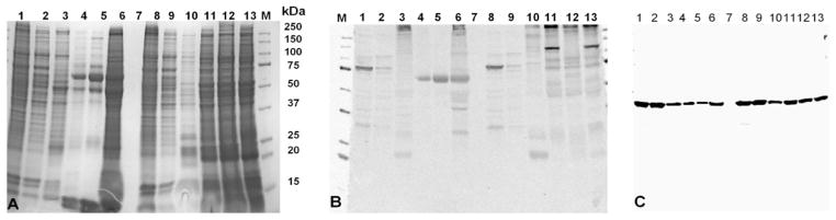
A 4–20% SDS-PAGE stained with GelCode Blue (A) and its corresponding immunoblot demonstrating cross-reactivity to the R. parkeri antibody (B) and β-actin (C). Standard protein marker adjoining molecular size lane (M); Lanes 1 and 2 were R. parkeri grown in Vero cells and the corresponding cell supernatant (Lanes 8 and 9). Lane 7 was empty. Lanes 3 and 10 were A. maculatum (Texas A&M) midgut and salivary gland tissues, respectively (Rickettsia-free tissues); Lanes 4 and 5 (midgut tissues) and lanes 11 and 12 (corresponding salivary glands), respectively, from field-collected A. maculatum; Lane 6 (midgut) and lane 13 (salivary gland) of lab colony A. maculatum (infected with R. endosymbiont).