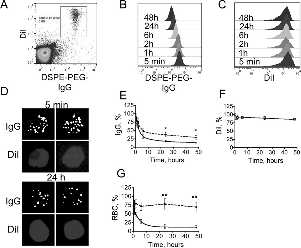
Fig. 2. In vivo stability of surface painted RBCs.
RBCs were painted with DiI and DSPE-PEG-IgG. Presence of DiI enabled to monitor RBCs independently of IgG. DSPE-PEG-IgG/DiI painted RBCs were injected into mice and the presence of the ligand was analyzed in blood samples taken at different times post-injection (after detecting IgG on RBCs with secondary antibody). A, Flow cytometry dot-plot of the injected RBCs. Double painted RBCs were easily distinguishable from majority of unlabeled cells; B, IgG FL-1 channel histogram shows significant shift at 24 h and 48h, suggesting gradual loss of IgG from the membrane; C, DiI fluorescence appears to be stable over the same time period; D, examples of individual double painted stained RBCs in the blood smear 5 min and 24 h post-injection. Contrast and brightness of all images were adjusted to the same degree; E, RBCs were prepared with different levels of DSPE-PEG-IgG painting (dotted trace–“low IgG”; solid trace–“high IgG”, see Result section). The clearance curve was fitted into bi-exponential decay with Prism software. There was significantly more retained IgG in case of “low IgG” painted RBCs (p-value 0.03, n=4, two-sided t-test). Low-IgG had a terminal half-life in the membrane of 74h; F, DiI on the same RBCs shows remarkable stability over time. DiI clearance could not be fitted into the exponential curve therefore connecting line is shown instead; G, RBC clearance is much lower for “low IgG” painted RBCs (dotted trace) than for “high IgG” painted RBCs (solid trace) (p-value 0.006, n=4, two-sided t-test).

