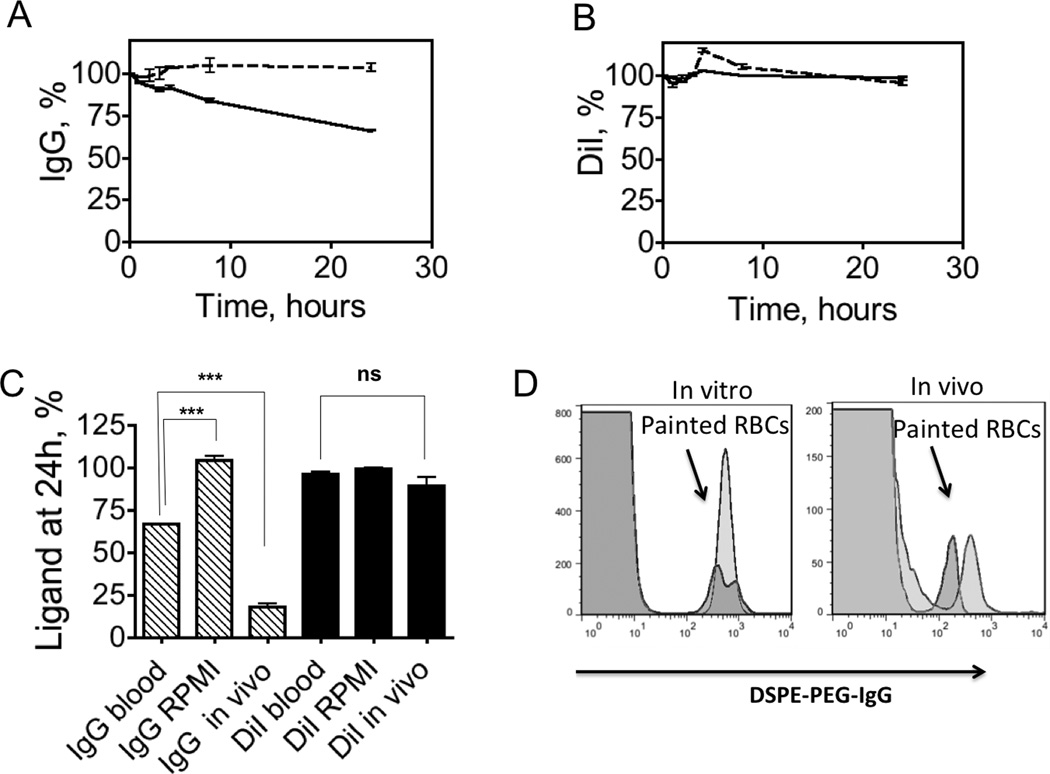
Fig. 3. In vitro vs. in vivo stability of surface painted RBCs.
RBCs were double painted as in Fig. 2 (“high IgG” painting) and stability of DSPE-PEG-IgG/DiI was monitored with flow cytometry (Supplemental Fig. S4). A, Stability of IgG and after incubation in full medium (dotted trace) and whole blood (solid trace). IgG levels are stable in medium, but less stable in whole blood over 24h incubation period; B, Stability DiI after incubation in medium (dotted trace) and full mouse blood (solid trace); C, Summary of IgG fluorescence and DiI fluorescence loss at 24h. Some IgG is lost in vitro due to exchange with blood components judging by the significant difference between ROMI and blood incubation levels (p-value<0.001, n=3, two-sided t-test). In vivo specific loss contributes significantly since there is a difference between IgG levels in blood in vitro and in vivo (p-value<0.001, n=3, two-sided t-test); D, Flow cytometry histograms of DSPE-PEG-IgG levels on RBCs in blood in vitro (left) and in vivo (right). Red trace is 5 min post-incubation (injection), blue trace is 24h post-incubation (injection). The histogram shows that there is no transfer of fluorescence from painted RBCs (arrow) to normal RBCs (major population) both in vitro and in vivo.

