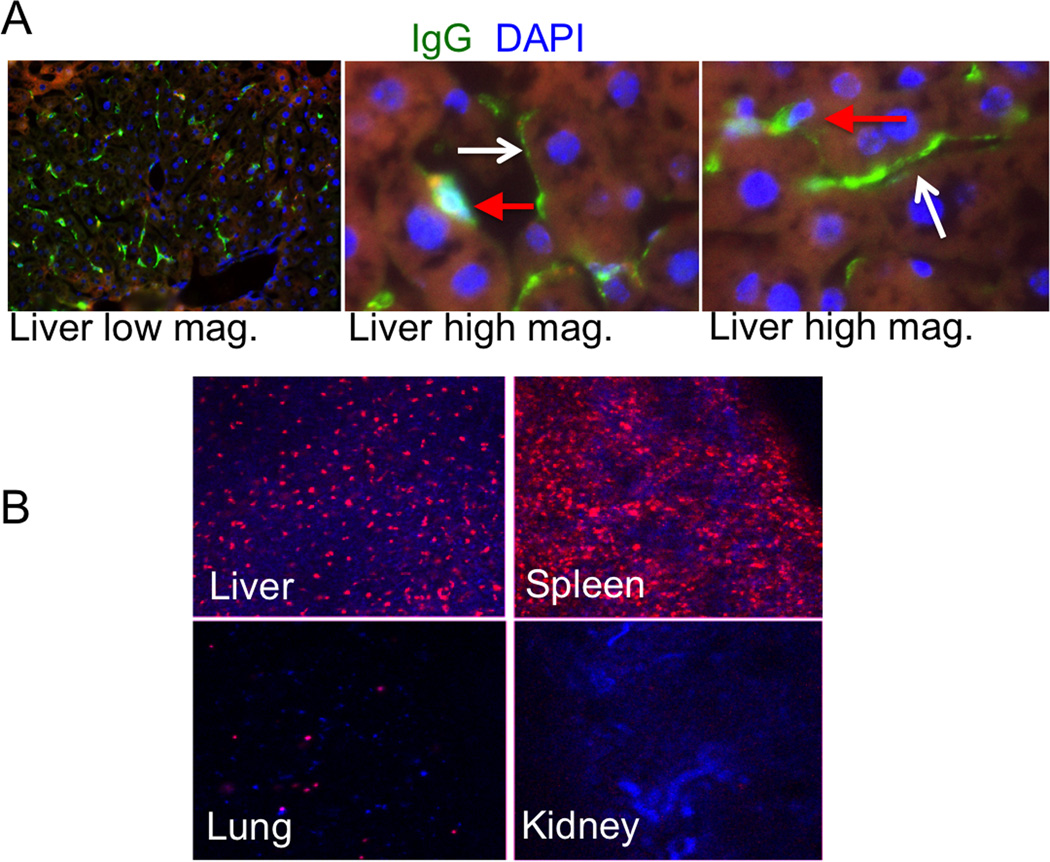
Fig. 4. Fate of surface IgG and painted RBCs in vivo.
A, Mice were injected with DSPE-PEG-IgG (rat) and the tissues were stained with Alexa 488 goat-anti-rat IgG. Liver tissue showed presence of DSPE-PEG-IgG stain. Left image is low magnification; center and right images are high magnification of the liver tissue. Red arrows point to depositions of rat IgG in Kupffer cells and white arrows point to rat IgG on endothelial (sinusoidal) cells; B, Mice were injected with DSPE-PEG-IgG/DiI painted RBCs. Low-magnification images of non-fixed tissue sections of mouse organs were taken 24h post-injection. Majority of DiI accumulated in the liver and spleen and trace amounts were found in lungs and kidneys.

