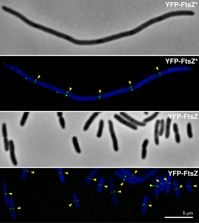
Figure 6. Localization of YFP-FtsZ and YFP-FtsZ* in FtsZ-depleted VIP2 cells.

Phase-contrast and fluorescence merged images of DAPI staining and YFP-fluorescence signal are shown for VIP2 cells producing YFP-FtsZ or YFP-FtsZ* at 42 °C after FtsZ depletion. Each sample was stained with DAPI to evidence bacterial nucleoids. Arrowheads mark the position of the FtsZ-rings.
