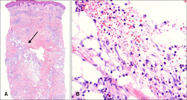
Fig. 2.

(A) A cavity with surrounding dense inflammatory cells infiltration and amorphous eosinophilic material (arrow) in the dermis (H&E, ×20). (B) Inflammatory cell infiltration composed of lymphocytes, neutrophil, and eosinophils in the dermis (H&E, ×400).
