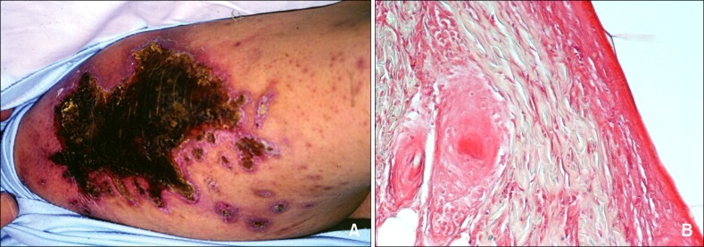
Fig. 1.

(A) Clinical appearance of the patient and (B) histopathologic examination of the cutaneous necrosis (H&E, ×200): skin necrosis with evidence of fibrin deposits in the post capillary venules, absence of arteriolar thrombosis, and lack of vascular or perivascular inflammation.
