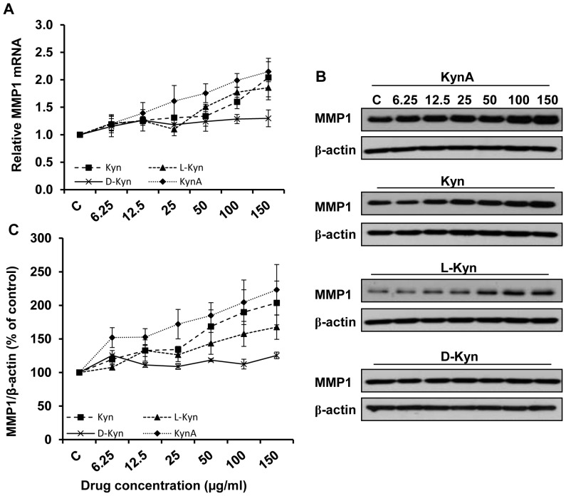
Figure 2. Stimulatory effect of kynurenines on MMP1 expression.
A: Dermal fibroblasts were treated with increasing doses (6.25, 12.5, 25, 50, 100, and 150 μg/ml) of KynA, Kyn, L-Kyn or D-Kyn. Following 24 hours of treatment cells were collected, and MMP1 expression was determined by Q-PCR after RNA extraction and cDNA synthesis. B: Evaluation of MMP1 expression at the protein level by Western blotting after 48 hours of treatment. C: The Mean±SEM ratio of MMP1 to β-actin density at the protein level. β-actin and GAPDH were used as loading controls for western blotting and Q-PCR, respectively.

