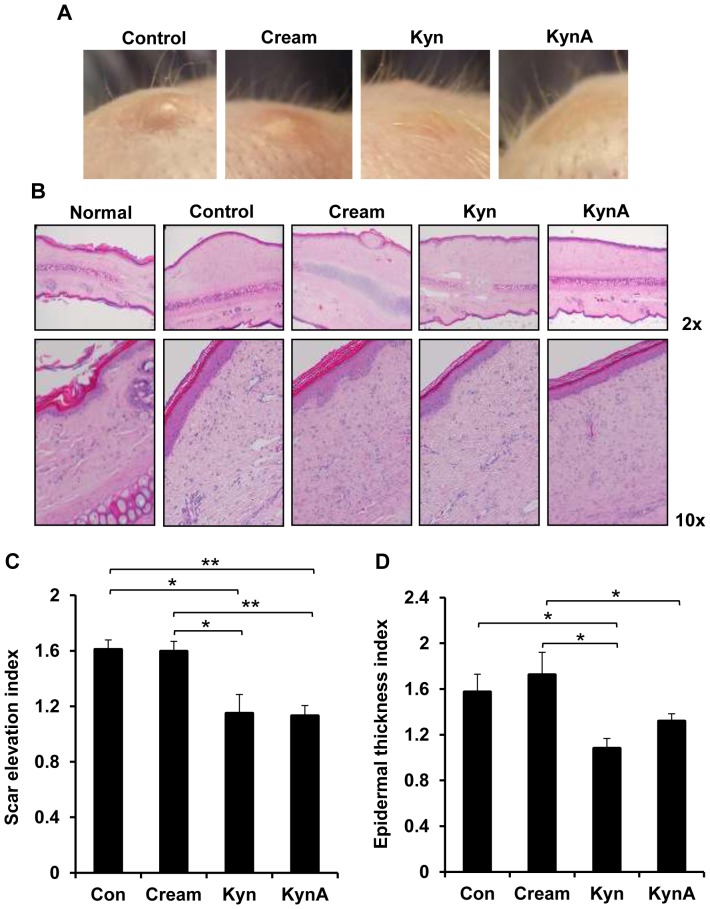
Figure 7. Clinical appearance and histological evaluation of wounds in rabbit ear model.
A: The clinical appearance of wounds that either received nothing (Control), cream only (cream) or cream containing Kyn or KynA on day 35 post wounding. B: Tissue samples were subjected to H&E staining to determine the dermal and epidermal hypertrophy. Scar elevation index (C) and epidermal thickness index (D) was evaluated quantitatively. Uninjured rabbit ear skin was used as the normal sample (* P-value<0.05 and ** P-value<0.01, n = 4).

