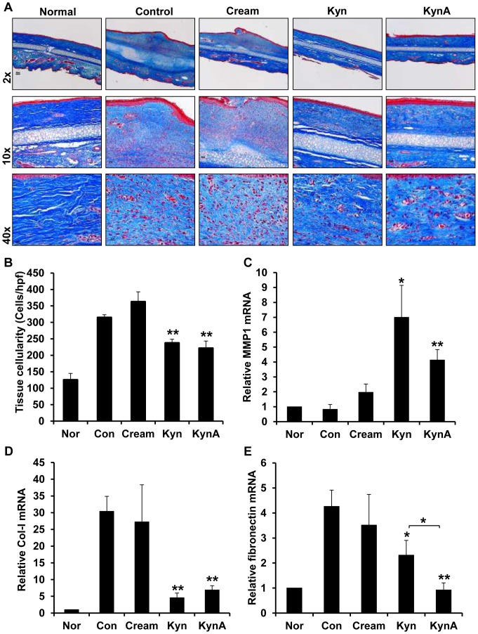
Figure 8. Effect of Kyn and KynA topical application on collagen deposition, tissue cellularity and ECM expression.
A: Evaluation of collagen deposition in tissue samples using Masson's Trichrome staining at day 35 post-wounding. In this staining collagen fibers are stained blue, keratin and muscle fibers are stained red, and cell cytoplasm and nuclei are stained light pink and dark brown, respectively. B: Quantification and statistical analysis of tissue cellularity. Q-PCR analysis of relative MMP1 (C), type-I collagen (D) and fibronectin (E) mRNA expression in tissue samples (* P-value<0.05 and ** P-value<0.01, n = 4).

