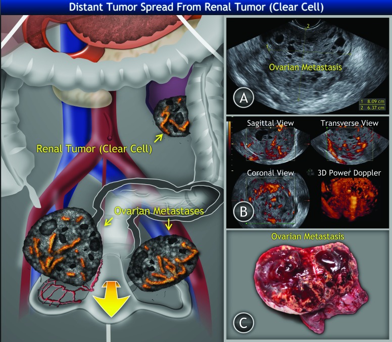
Fig. 1.
Clear cell renal cell tumour of the left kidney with bilateral ovarian metastases: A: Ultrasound findings of right ovarian metastasis of solid structure with multiple small irregular cysts filled with hypoechogenic (blood) intracystic fluid. B: Three-dimensional power Doppler ultrasound of ovarian metastasis showing tumour structure and vessel tree. C: Gross appearance of the ovarian tumour.

