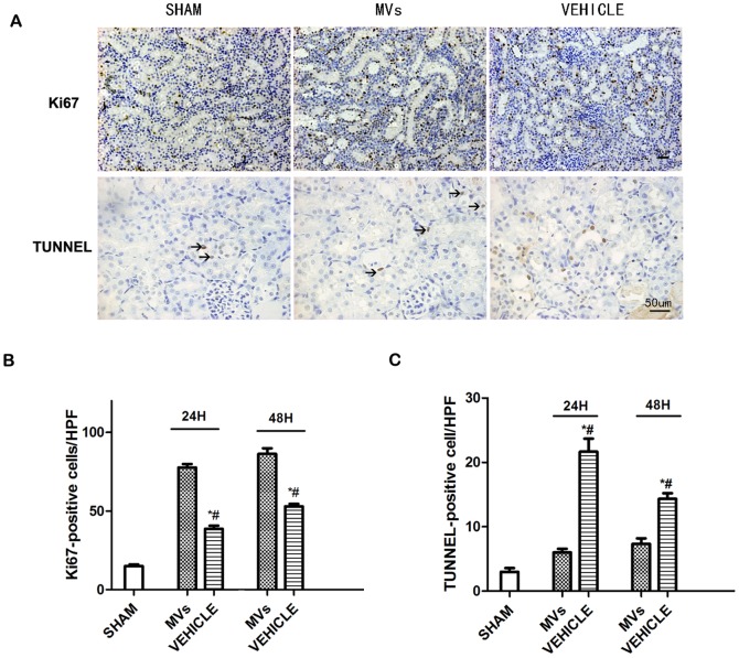
Figure 2. Renal cells apoptosis and proliferation in acute kidney injury rats.
(A) Representative micrographs of Ki67 (magnification ×200, Scale bar = 50µm) and TUNEL (magnification ×400, Scale bar = 50 μm) immunostaining in the injured kidneys at 48 h after reperfusion. (B) Quantification of TUNEL-positive tubular epithelium cells in the kidney sections at different time points after reperfusion (n = 6).*P<0.05, MVs versus VEHICLE; #P<0.05, SHAM versus VEHICLE. (C) Quantification of Ki67-positive tubular epithelium cells in the kidney sections at different time points after reperfusion (n = 6). *P<0.05, MVs versus VEHICLE; #P<0.05, SHAM versus VEHICLE.

