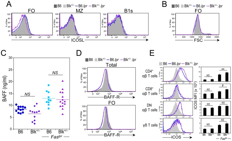
Figure 6. Evidence for augmented ICOS-ICOSL interactions in Blk+/−.lpr mice.
(A) Representative histograms showing ICOSL levels on follicular (FO), marginal zone (MZ) and splenic B1 (B1s) B cells from 3-month-old B6 (n = 6), Blk+/− (n = 6), B6.lpr (n = 7) and Blk+/−.lpr (n = 8) mice. (B) Histogram comparing B cell size, using FSC units, among 3-month-old B6 (n = 6), Blk+/− (n = 6), B6.lpr (n = 7) and Blk+/−.lpr (n = 8) mice. (C) Comparison of the BAFF serum levels in 3-month-old B6, Blk+/−, B6.lpr and Blk+/−.lpr mice. Each symbol represents an individual mouse. (D) Representative histograms comparing BAFF-R levels on total (CD19+) (top) and FO B cells (bottom) from 3-month-old B6 (n = 8), Blk+/− (n = 10), B6.lpr (n = 13) and Blk+/−.lpr (n = 12) mice. (E) Representative histograms showing the expression of ICOS on CD4+, CD8+, DN αβ, and γδ T cells subsets from the spleens of 3-month-old B6.lpr and Blk+/−.lpr mice. ICOS levels on the corresponding T cell subsets from age-matched B6 mice are also shown (shaded histogram). Adjacent graph compares ICOS levels (MFI) on each T cell subset between 3-month-old B6 (n = 6) and Blk+/− (n = 6) mice and between 3-month-old B6.lpr (n = 7) and Blk+/−.lpr (n = 8) mice.

