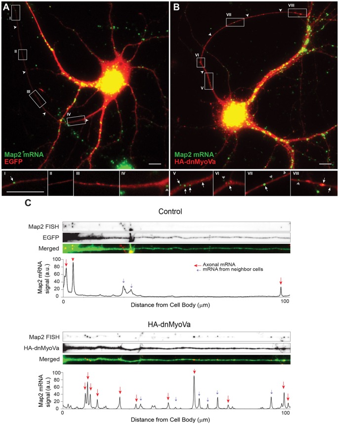
Figure 3. Myosin Va is necessary for somatodendritic targeting of endogenous Map2 mRNA.
A. Map2 mRNA labeled by fluorescent in situ hybridization (green) in a cortical neuron in dissociated culture expressing EGFP (red). Map2 mRNA is present in the somatodendritic compartment, but largely absent from the axon (arrowheads). Insets I-IV are high power images corresponding to boxes drawn in (A). B. In a cortical neuron expressing HA-dnMyoVa, staining for endogenous Map2 mRNA is present in the axon as well as in the somatodendritic compartment. Insets V-VIII are high power images corresponding to boxes in (B). Arrowheads point to axons, arrows to Map2 mRNAs that are present in the axon, double arrowheads to Map2 mRNAs from neighboring untransfected neurons. Scale bar, 10 μm. C. Signal intensity plots of Map2 mRNA staining in straightened axons of the control neuron in (A) and the neuron expressing HA-dnMyoVa in (B). The presence of HA-dnMyoVa is correlated with an increase in the amount of Map2 mRNA in the axon. Red arrows point to Map2 mRNA puncta in the axon. Blue arrows point to Map2 mRNA puncta from neighboring neurons.

