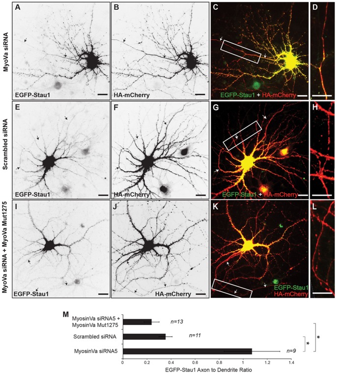
Figure 4. Knockdown of Myosin Va disrupts somatodendritic targeting of EGFP-Stau1.
A. EGFP-Stau1 localizes to both the axon and the somatodendritic compartment in a cortical neuron transfected with MyoVa siRNA and HA-mCherry (B). C. Merge of EGFP-Stau1 (green) and HA-mCherry (red). D. High power image of boxed area in (C). E. EGFP-Stau1 localizes in the soma and dendrites, but not in the axon, of a neuron expressing scrambled siRNA and HA-mCherry (F). G. Merge of EGFP-Stau1 (green) and HA-mCherry (red). H. High power image of boxed area in (G). I. EGFP-Stau1 localizes in the soma and dendrites, but not in the axon, of a neuron expressing MyoVa siRNA as well as MyoVa Mut1275 and HA-mCherry (J). K. Merge of EGFP-Stau1 (green) and HA-mCherry (red). L. High power image of boxed area in (K). Scale bar, 10 μm. Note that this result indicates that blocking of somatodendritic localization of EGFP-Stau1 by MyoVa siRNA is not due to off-target effects. M. Axon to dendrite ratio of EGFP-Stau1 in neuron expressing MyoVa siRNA is significantly different from those in neurons expressing either scrambled siRNA or MyoVa siRNA and MyoVa Mut1275. * indicates p<0.001 (Kruskal-Wallis).

