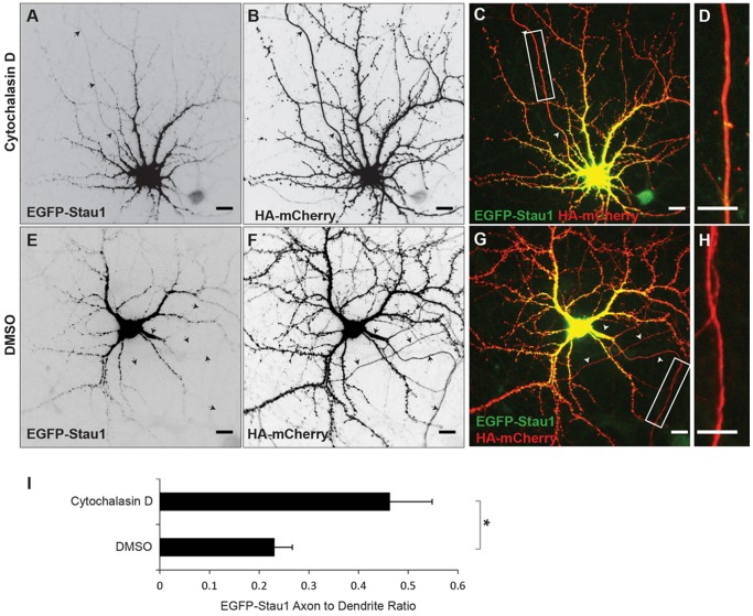
Figure 5. Intact actin filaments are necessary for somatodendritic localization of EGFP-Stau1.
A. Following exposure to Cytochalasin D (4 μM) for 3 hours EGFP-Stau1 is present in the axon as well as in the dendritic compartment. B HA-mCherry staining in the same cell as in A. C. Merge of EGFP-Stau1 (green) and HA-mCherry (red). D. High power image of boxed area in (C). E. EGFP-Stau1 is absent from the axonal compartment of cortical neurons expressing HA-mCherry (F) and exposed to DMSO. G. Merge of EGFP-Stau1 (green) and HA-mCherry (red). H. High power image of boxed area in (G). I. EGFP-Stau1 axon to dendrite ratio is significantly higher in cells exposed to Cytochalasin D than in cells exposed only to DMSO. * indicates p<0.03 (Wilcoxon). Scale bar 10 μm.

