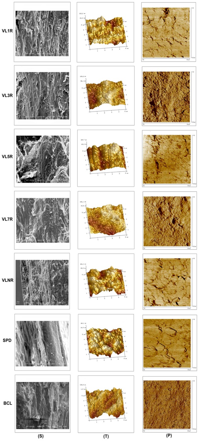
Figure 5. Scanning electron microscopy (SEM) images (Mag = 500X), AFM topographic images (5 μm×5 μm) and phase images (5 μm×5 μm) of the trabecular bone in femoral head.

Sample thickness is 2 mm. Column (S): scanning electron microscopy; Column (T): AFM topographic images; Column (P): AFM phase images. Row VL1R: vibrational loading for 1 d followed by 1 d rest group; Row VL3R: vibrational loading for 3 d followed by 3 d rest group; Row VL5R: vibrational loading for 5 d followed by 5 d rest group; Row VL7R: vibrational loading for 7 d followed by 7 d rest group; Row VLNR: vibrational loading for no rest day or vibrational loading every day; Row SPD: tail suspension group; Row BCL: baseline control group.
