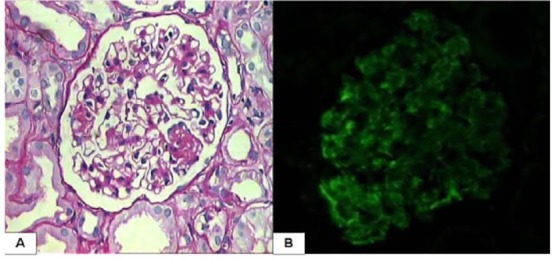
Figure 1.

Two representative pictures, one of light microscopy and other of immunofluorescence from a renal biopsy from an 8 year old child with steroid dependant nephrotic syndrome. The interpretation of these findings is open to all the readers. A. This is high-power view of one representative glomerulus from the above case. (PAS, ×400). B. Immunofluorescence staining for IgM by the direct technique on the same biopsy. (IgM, ×400).
