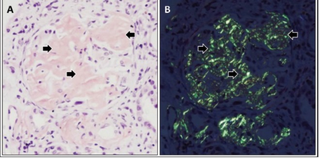
Fig. 1.

Renal amyloid A amyloidosis. A photomicrograph demonstrating accumulation of amorphous material (arrows)in a glomerulus (A). Polarizing microscopy revealing apple-green birefringence of deposits in the glomerular mesangial areas (arrows) (B). Tissue sections were stained with Congo red. Courtesy of David B. Thomas, MD.
