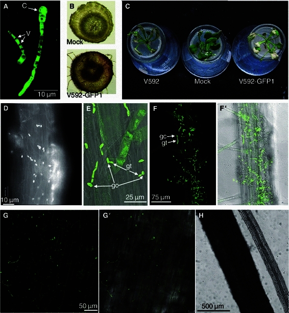
Fig. 1.

Infectivity of V V592-GFP1 and confocal microscopy images of the initial stage root colonization by V592-GFP1 on cotton and Arabidopsis roots. (A) Germinating conidium and hyphae of V592-GFP1. The arrows indicate vacuoles of fungal cells visible as dark areas in the fluorescing cytoplasm. C, conidium, V, vacuole. (B) V592-GFP1-infected cotton stem vascular displayed discoloration compared to mock inoculation. (C) Infectivity of V592-GFP1 relative to V592. Similar leaf wilt symptoms were observed with V592-GFP1 and V592 on Arabidopsis. Photographs were taken two weeks post-inoculation. Mock, mock-inoculation. (D) Conidia-covered Arabidopsis root 6 h post-inoculation (hpi). (E) Germ tubes emerging from one end of the conidium (12 hpi). gc, germinated conidia, gt, germ tubes. (F) Fluorescence image of V592-GFP1 hyphae on a root of Arabidopsis around the root hair zone (24 hpi). (F′) Compound micrograph of bright field transmission and corresponding fluorescence images (same view as F). (G) Fluorescence image of V592-GFP1 hyphae on a root of cotton (24 hpi). Image displays some auto-fluorescence of cotton root epidermal cells. (G′) Compound micrograph of bright field transmission and corresponding fluorescence images (same view as G). (H) Comparison of cotton and Arabidopsis roots under bright field transmission micrograph
