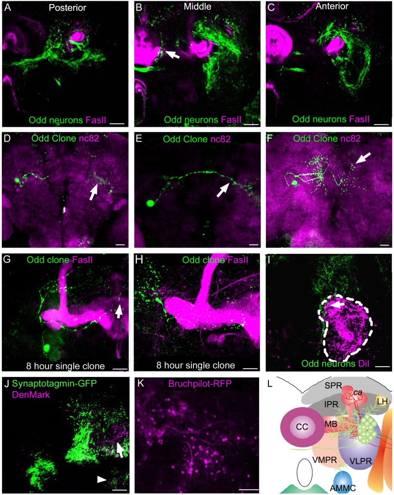
Figure 6.
Characterization of the Odd projections into the IPR, VMPR, VLPR and PLPR. All images are posterior views of adult brains, dorsal up. A–C: Single optical sections (×40) at three different anterior–posterior levels at the level of the MB lobes. The Odd neurons appear to wrap around the MB lobes and also project toward the central complex (arrow B). D–F: Low-magnification (×20) maximum intensity stack images of single-cell MARCM Odd clones (green) in brains stained with nc82. D: Image composed of 10 sections (total depth 20 μm), showing an Odd MARCM single-cell clone induced at second instar larval stage. This neuron projects into the same compartment (VMPR) in each brain hemisphere (arrow). E: Image composed of eight sections (total depth 16 μm) showing an Odd single-cell MARCM clone induced at late second larval stage. This neuron projects into the same area within the IPR in each brain hemisphere (arrow). F: Image composed of eight sections (total depth 16 μm), showing a single-cell MARCM Odd clone induced at 7–8 hours of embryonic development. This cell projects to different areas within the same compartment in both brain hemispheres (arrow). G,H: Images (×40) of single-cell MARCM clones (green) with the neuropil stained with the FasII antibody (magenta) to label the MB. G: Image composed of eight confocal sections (total depth 16 μm). This clone was induced at first instar larval stage and shows a neuron that projects to different parts of the VMPR within the same hemisphere. H: Image composed of nine sections (total depth 18 μm). Single-cell clone that projects to the IPR and the VMPR within the same brain hemisphere. I: Life image (×40) of Odd neurons colabeled with DiI retrogradely labeled neurons (magenta) projecting into the AMMC (dashed lines). The majority of the Odd neurons lie posterior and dorsal to the AMMC, although there are a few Odd projections at the dorsal aspect of the AMMC (arrow).This image is a maximum intensity stack composed of 16 sections (total depth 32 μm). J: Coexpression of synaptotagmin-GFP and DenMark in the Odd neurons. This maximum intensity projection image (×20) is composed of six sections (total depth 12 μm). Although the majority of neurites in the IPR, VMPR, VLPR, and PLPR compartments are axonal, dendrites are also present in particular in the PLPR region (arrows). K: Another axonal marker; Bruchpilot-RFP (magenta), was used to confirm the axonal identity of the IPR, VMPR, VLPR, and PLPR arbor. This image is ×40 magnification and a maximum intensity stack composed of five sections (total depth 10 μm). L: Schematic representation of the Odd axonal (green) and dendritic (magenta) projections. SPR, superior protocerebrum; IPR, inferior protocerebrum; ca, calyx; MB, mushroom body; CC, central complex; LH, lateral horn; VMPR, ventromedial protocerebrum; VLPR, ventrolateral protocerebrum; AMMC, antennal mechanosensory and motor center. Scale bar = 50 μm in A–K. [Color figure can be viewed in the online issue, which is available at wileyonlinelibrary.com.]

