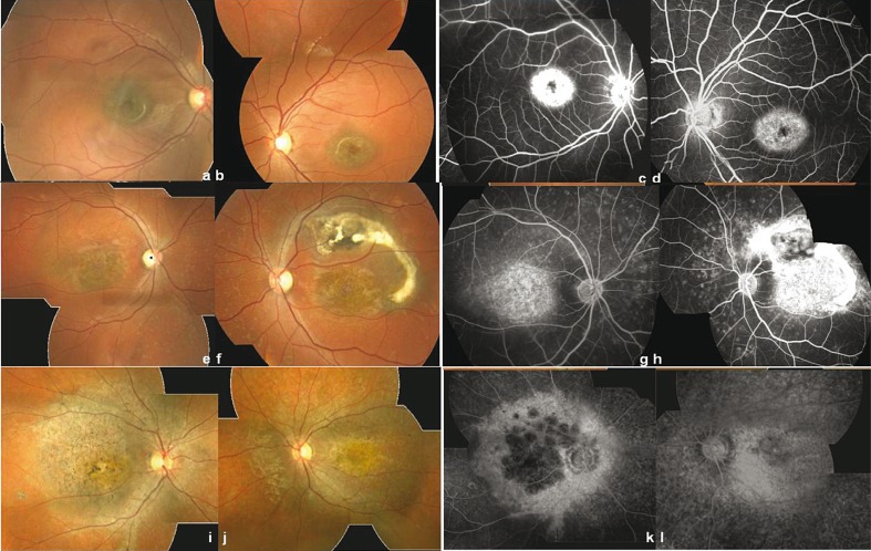
Figure 2.
Polymorphic expression of Stargardt disease in family A based on funduscopy and angiography. Typical Stargardt with bull’s eye maculopathy; fundus appearance (a, b) and angiograms (c, d) in phenotype I (right eye results: a, c; left eye results: b, d). Fluorescein angiography shows the typical appearance of reduced transmission of background fluorescence (dark choroid) in patient V-7. Stargardt disease associated with fundus flavimaculatus; fundus appearance (e, f) and angiograms (g, h) in phenotype II (right eye results: e, g; left eye results: f, h). Composite image of the right eye shows flecks throughout the posterior pole, atrophic macular changes and several flecks. Patient V-6 also displays a fibroglial scar in the left eye (f, h). Fluorescein angiography clearly reveals the dark choroid. Advanced stage of Stargardt disease or cone rod dystrophy; fundus appearance (i, j) and angiograms (k, l) in phenotype III (right eye results: i, k; left eye results: j, l ). Patient V-1 presents a large demarcated atrophic area in the macula with pigment clumping and migration extending to the peripheral retina illustrating an overlapping phenotype.

