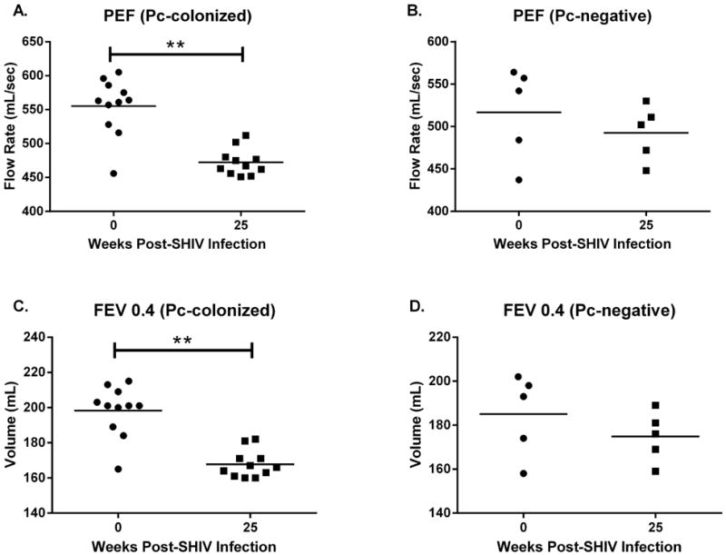
Figure 3. Pneumocystis-colonized macaques exhibited significant declines in pulmonary function, whereas pulmonary function was preserved in Pneumocystis-negative animals.

Significant declines in both peak expiratory flow (PEF, p=0.001, A) and forced expiratory volume in 0.4s (FEV 0.4, p=0.001, C) occurred in Pc-colonized macaques (n=11, paired t-test, week 0 vs week 25 post SHIV-infection). At 25 weeks post SHIV-infection, PEF (B, p=0.21) and FEV 0.4 (D, p=0.21) were similar to baseline values (n=5, paired t-test) in the Pc-negative animals.
