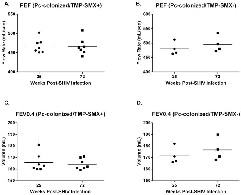
Figure 5. Pulmonary function remained steady in both TMP-SMX-treated and untreated Pneumocystis-colonized macaques for the remainder of the study (weeks 25-72 post SHIV-infection).

Peak expiratory flow (PEF, p=0.29, A) and forced expiratory volume in 0.4s (FEV 0.4, p=0.46, C) values were similar at experiment termination (72wpi), compared with pulmonary function measurements taken prior to TMP-SMX initiation (25wpi), in the TMP-SMX-treated macaques. PEF (B, p=0.39) and FEV 0.4 (D, p=0.39) values in the Pc+, untreated group were also not different compared to the time-point at which TMP-SMX was initiated in the treatment group (paired t-test). PEF and FEV 0.4 did not decline significantly by 72wpi in the Pc-negative group (data not shown, p=0.70).
