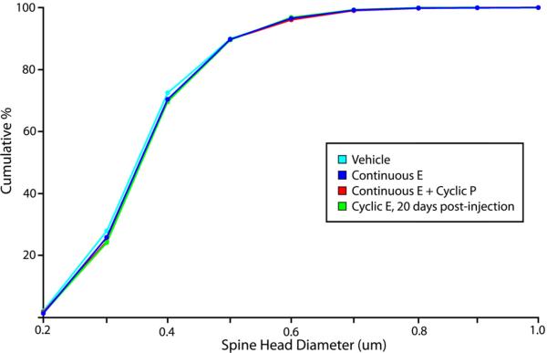
Figure 3. Dendritic spine morphology in area 46.
Previous results showed that, 24 hours after injection, cyclic E administration in both young OVX primates led to a left shift in the cumulative distribution curve for spine head diameter, indicating a greater proportion of thinner spines in the E-treated animals (Hao et al., 2007). We found that continuous E treatment, with or without cyclic P, does not increase the proportion of thinner spines in the dlPFC of young OVX animals, despite reaching similar levels of circulating E. We also found that, 20 days after the final E injection in the context of long-term cyclic E replacement, the distribution of spine head diameters in the dlPFC is not significantly different from that seen in vehicle-treated animals.

