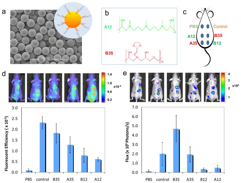
Figure 3.
Imaging of live animals with subcutaneously-injected PBAA-coated microparticles. (a) SEM image of carboxylated polystyrene beads used as the substrate for PBAA coatings. The inset is the schematic of the PBAA coating on the particles (not to scale). (b) Chemical structures of A12 and B35, which inhibits and promotes the activation of monocyte/macrophage cells in vitro, respectively. (c) The configuration of 6 injections on the back of a mouse. (d) Fluorescent images of mice (5 replicates) 24 hours after injections using Prosense as the probe and quantitative results from the image analysis. (e) Luminescent image of the same 5 mice using Luminol as the probe and quantitative results.

