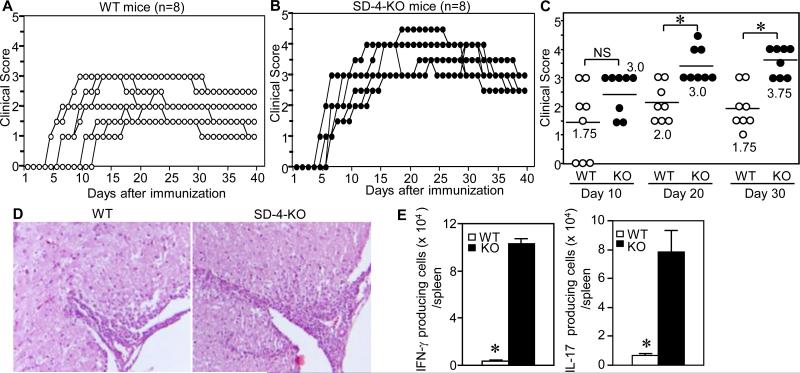
Figure 2. SD-4−/− mice develop significantly worse EAE.
WT (A) or SD-4 KO (B) mice (n=8) were immunized with MOG peptide and complete Freund's adjuvant on day 0 and boosted with pertussis toxin on days 0 and 2. Kinetics of EAE development were monitored by clinical score of each mouse, plotted in a graphic version. Clinical score: 0, no abnormality; 1, flaccid tail; 2, moderate hind limb weakness; 3, severe hind limb weakness; 4, complete hind limb paralysis; and 5, quadriplegia, moribund state. (C) Clinical scores in early- (day 10), mid- (day 20) or late-phases (day 30) were plotted on a scatter chart (median, n=8), with statistical significance of scores (ANOVA, *p<0.001; NS, not significant) between WT and KO mice. (D) Spinal cords of mice 20 d after immunization were stained with H&E and examined under a microscope (4 X magnification). (E) IFN-γ- or IL-17-producing cells in spleen of WT and KO mice 10 d after immunization were enumerated by ELISPOT assay and calculated as number per spleen. *p<0.001 between WT and KO mice. All data shown are representative of 3 separate experiments.

