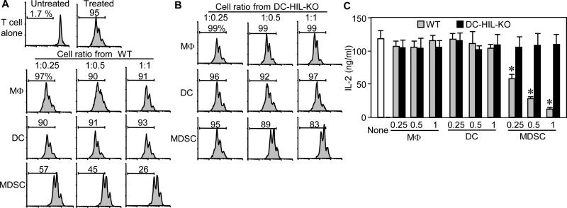
Figure 5. Among DC-HIL-expressing myeloid cells in EAE-developing mice, MDSC are the most potent suppressors.
(A and B) Three myeloid cell subsets were purified from spleen of WT (A) or DC-HIL KO (B) mice that were EAE-immunized 10 days prior. These cells were assayed separately for T cell-suppressive activity by coculturing with CFSE-labeled WT-T cells at varying cell ratios in the presence of anti-CD3/CD28 Ab. Three days after culture, % of proliferated cells was examined by flow cytometry and data are shown in histograms. (C) Similar assays were performed, but with unlabeled T cells. T cell activation is shown by IL-2 production (mean ± sd, n=3). “None” means T cells and Ab without myeloid cells. *p<0.001 between WT and KO mice. Data are representative of 3 independent experiments.

