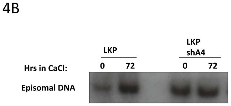
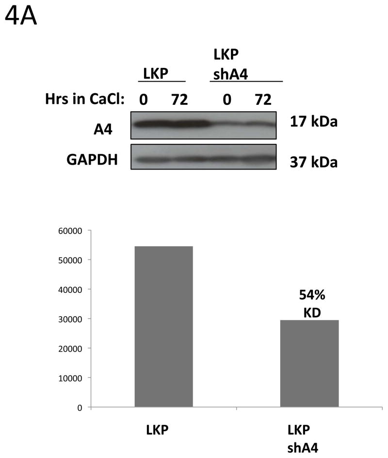
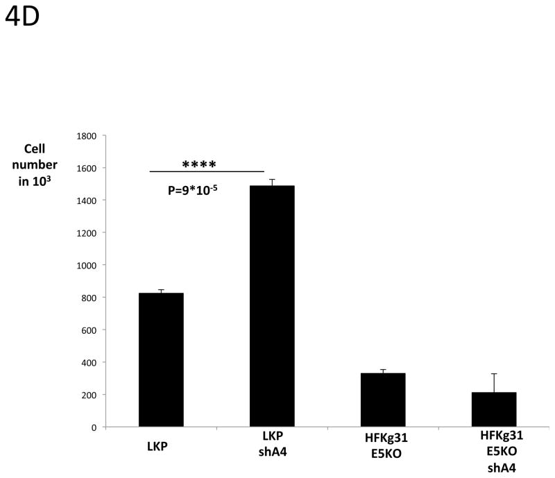
Figure 4. HPV31 replication and cell proliferation are enhanced in A4 shRNA knock down keratinocytes.


4A: Western blot analyses of A4 stable knock down in keratinocytes.
HPV31 positive keratinocytes LKP were infected with lentiviral particles carrying shRNAs against the A4 gene. Infected cells were selected with puromycin for two weeks. ImageJ program was used to quantify knockdown of A4 in cell line LKPshA4.
4B: Southern blot analysis of LKP and LKPshA4 keratinocytes.
Total genomic DNA was harvested from monolayer and differentiated cultures and digested with DpnI to remove residual input DNA and XbaI to linearize the HPV31 genomes. The Southern blot was hybridized with a probe that contains the complete HPV31 genome.
4 C, D: Cell proliferation assay.
LKP, LKPshA4, HFKg31E5KO and HFKg31E5KOshA4 cell lines were differentiated by suspension culture in methylcellulose. 48 hours later cells were re-plated into new dishes and cultured for few days. Colonies were visible under microscope on day 5 (4C). The total cell number was estimated on day 5 using TC10 automated cell counter (BioRad) (4D). Data are means ± standard deviations (error bars), n=3. ****P < 0.0001 indicates values significantly different from the control (LKP).


