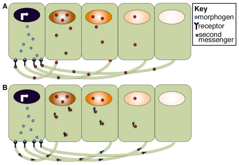
Figure 7. Model for cytoneme function in morphogen gradient formation.
Morphogen receiving cells extend long actin-based filopodia toward morphogen secreting cells. Receptors are proposed to bind the morphogen at points of contact and to either activate second messengers that then traffic back to the body of the target cell (A) or to directly traffic the bound morphogen to receiving cells (B). Illustration is schematic and not meant to imply that morphogen movement is restricted to one side of the cell.

