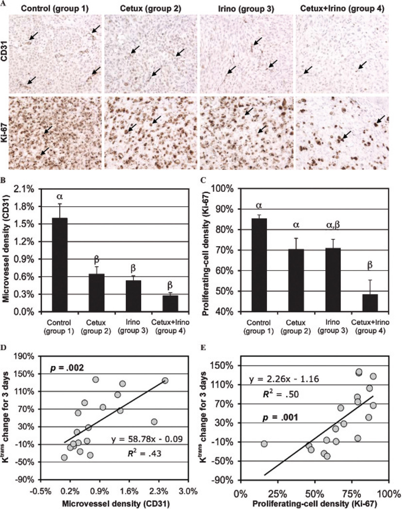
Figure 5.
A, Representative microphotographs of CD31 (×400 original magnification) and Ki-67 (×400 original magnification) staining of MIA PaCa-2 tumors of groups 1 to 4 collected at day 21; the microvessel areas and proliferating cells are indicated with black arrows in each row. Microvessel (B, CD31 expressed) and proliferating (C, Ki-67 expressed) cell densities of groups 1 to 4 are presented; statistical differences among groups are indicated by different Greek letters above bars. Ktrans changes in the peripheral tumor region of groups 1 to 4 during 3 days after therapy initiation versus (D) microvessel and (E) proliferating cell densities; p values represent the significance of the correlation.

