
Fig. 7.
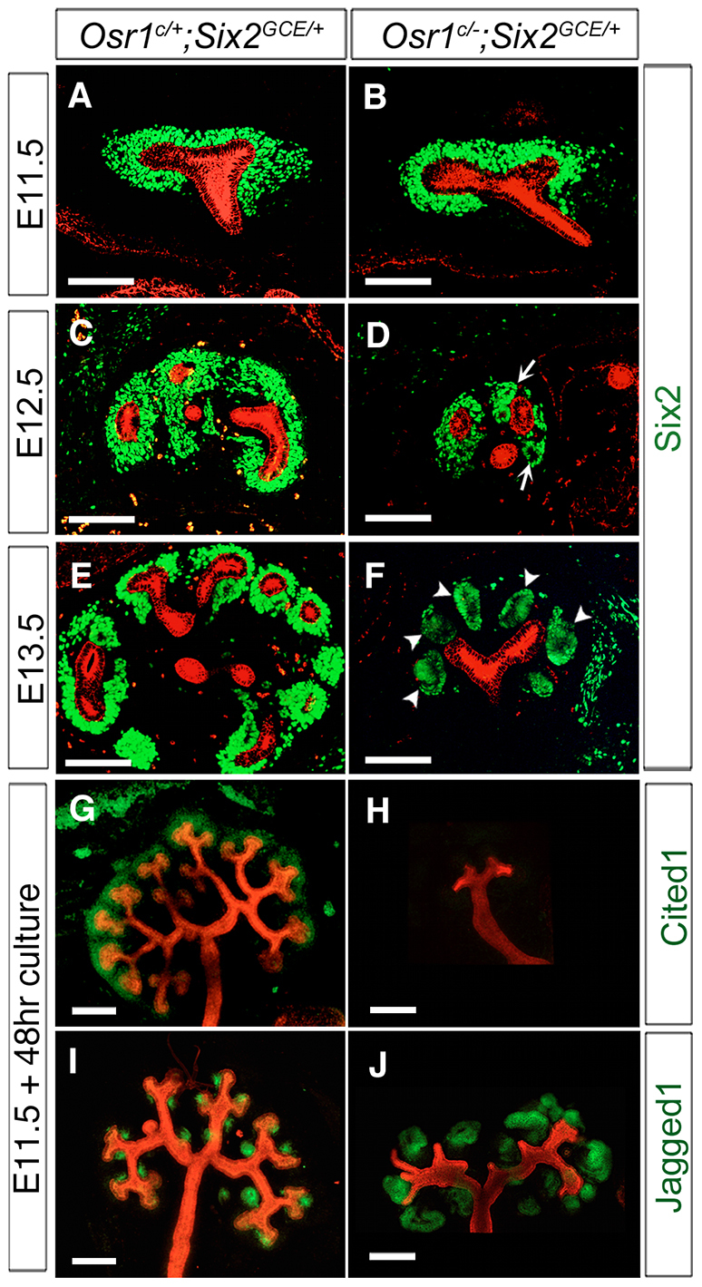
Analysis of cap mesenchyme and UB branching in the Osr1c/-;Six2GCE/+ mutant embryos. (A-F) Immunofluorescence detection of Six2 (green) and pan-cytokeratin (red) in developing kidneys of Osr1c/+;Six2GCE/+ (A,C,E) and Osr1c/-;Six2GCE/+ (B,D,F) embryos at E11.5 (A,B), E12.5 (C,D) and E13.5 (E,F). Arrows in D indicate abnormal mesenchymal aggregates surrounding the UB in the E12.5 Osr1c/-;Six2GCE/+ mutant kidney. Arrowheads in F indicate large epithelializing aggregates in place of the CM surrounding the UBs. Scale bars: 100 μm. (G-J) Whole-mount immunofluorescence detection of Cited1 (green, G,H) or Jag1 (green, I,J) and pan-cytokeratin (red) in cultured Osr1c/+;Six2GCE/+ (G,I) and Osr1c/-;Six2GCE/+ (H,J) kidney explants. Scale bars: 200 μm.