
Fig. 6.
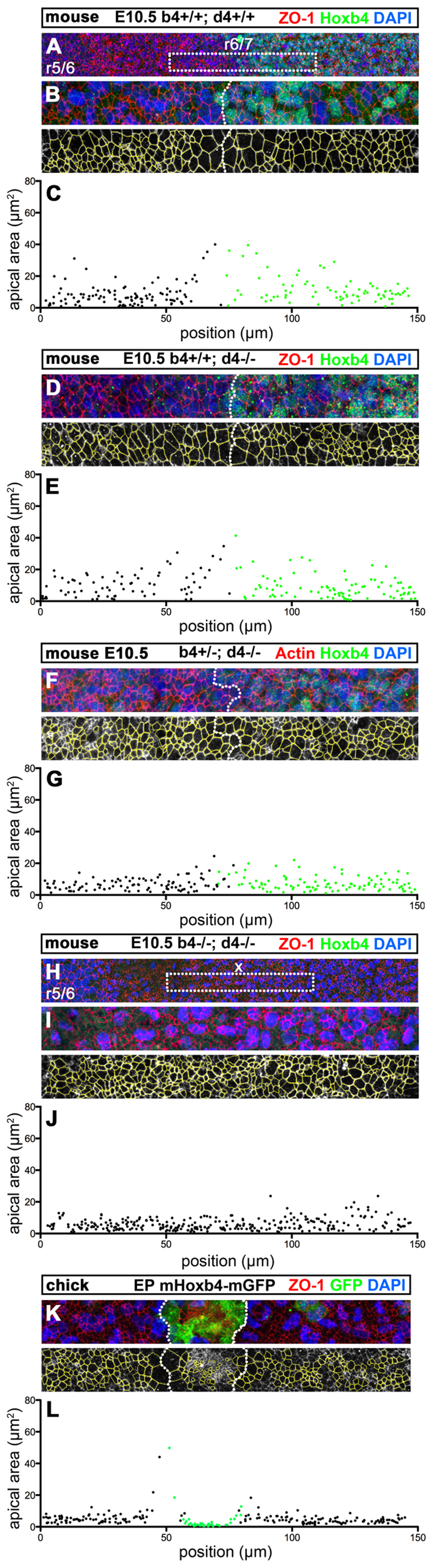
Hox4 proteins are necessary and sufficient to induce non-autonomous apical cell enlargement. (A-H) Confocal immunostaining for Hoxb4 (green) and ZO-1 (red) on E10.5 flat-mounted mouse hindbrains of wild-type (Hoxb4+/+; Hoxd4+/+) (A,B), Hoxb4+/+; Hoxd4-/- (D), Hoxb4+/-; Hoxd4-/- (F) and Hox4 double-mutant (H,I) embryos at a lateral level of the r6/r7 boundary region. The r6/r7 boundary or its presumptive position (x) are indicated in A and H. (B,I) Confocal z-projections of similar magnifications of the r6/r7 boundary region as in D (corresponding to dotted boxes in A and H) showing the apical cell outlines (yellow) used for quantitation. (C,E,G,J) Apical areas (μm2) with respect to the anteroposterior position of the cells in B,D,F,I, respectively. Hoxb4+ (green) and Hoxb4- (black) cells are indicated. (K,L) Confocal z-projections and ZO-1 apical outlines (yellow) of the chick r5 neuroepithelium electroporated with mouse Hoxb4-mGFP. (K) Mouse Hoxb4/GFP+ cells (green) form a cluster with a well-defined interface (dotted line). (L) Corresponding apical cell areas (μm2) with respect to anteroposterior position for mouse Hoxb4+ (green) and mouse Hoxb4- (black) cells. Anterior is towards the left.