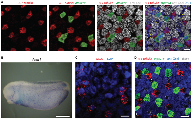
Fig. 1.
foxa1 marks a new epidermal cell type. (A) Double fluorescence in situ hybridisation and antibody staining for a ciliated cell marker (α-1-tubulin, red), an ionocyte marker (atp6v1a, green) and a goblet cell marker (anti-Xeel, grey) on stage 32 embryos. DAPI (blue) is used to mark each cell. At least one cell type (yellow circles) is not stained by these markers. (B) Chromogenic in situ hybridisation for foxa1 on stage 25 embryos shows scattered, spotted epidermal distribution. (C) foxa1 is epidermally expressed in a subset of DAPI-positive cells. (D) foxa1 expression (grey) completes the epidermal staining when added to markers of the other cell types at stage 32. Scale bars: 20 μm in A,C,D; 500 μm in B.

