Fig. 2.
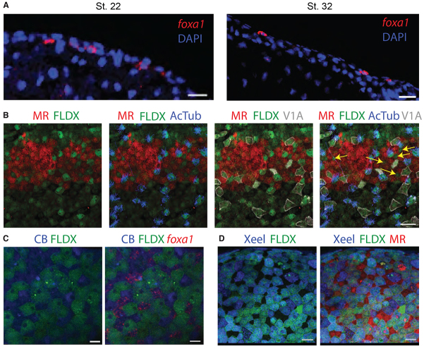
foxa1 cells intercalate from inner to outer layer at mid-tailbud stages. (A) Sections of stage 22 and stage 32 embryos stained for DAPI (blue) and foxa1 (red) by fluorescent in situ hybridisation shows that expression changes from inner to outer layer as the embryo develops. (B) Transplant of MR-labelled (red) outer layer epidermal tissue on to FLDX-labelled (green) host embryo and stained using antibodies for ciliated cell marker, acetylated α-tubulin (AcTub, blue) and ionocyte marker, V1a (grey) at stage 32. A cell type (yellow arrows) intercalates from inner to outer layer in addition to ciliated cells and ionocytes. (C) Transplant of FLDX-labelled (green) outer layer epidermal tissue on to CB-labelled (blue) host embryo and stained by fluorescent in situ hybridisation for foxa1 (red) at stage 32. (D) Transplant of FLDX-labelled (green) outer layer epidermal tissue on to MR-labelled (red) host embryo and stained with anti-Xeel antibody (blue). Scale bars: 50 μm in A,B,D; 15 μm in C.

