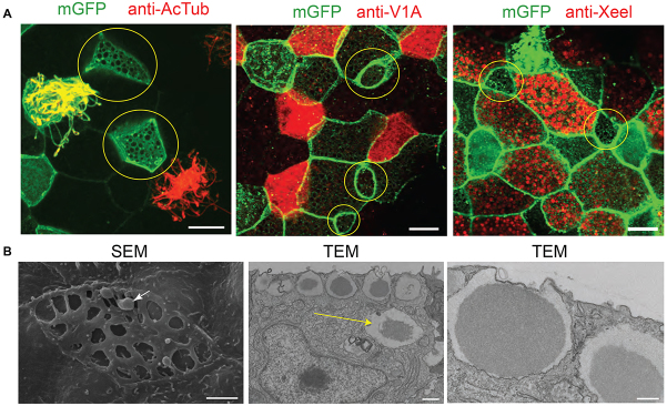
Fig. 3.

Small cells with large vesicles containing secretory material. (A) Small cells (yellow circles) are not labelled by markers for ciliated cells (anti-AcTub), ionocytes (anti-V1a) or goblet cells (anti-Xeel). Membranes are marked with membrane GFP (mGFP). (B) SEM shows small cells with large apical openings and secretory material highlighted with an arrow. Sections imaged by TEM show vesicles at the apical membrane containing a dark core surrounded by lighter material. Highlighted with a yellow arrow is a vesicle deeper within the cytoplasm that may represent an immature vesicle. Scale bars: in A, 10 μm; in B, 2 μm (SEM), 1 μm (TEM, low magnification) and 0.5 μm (TEM, high magnification).
