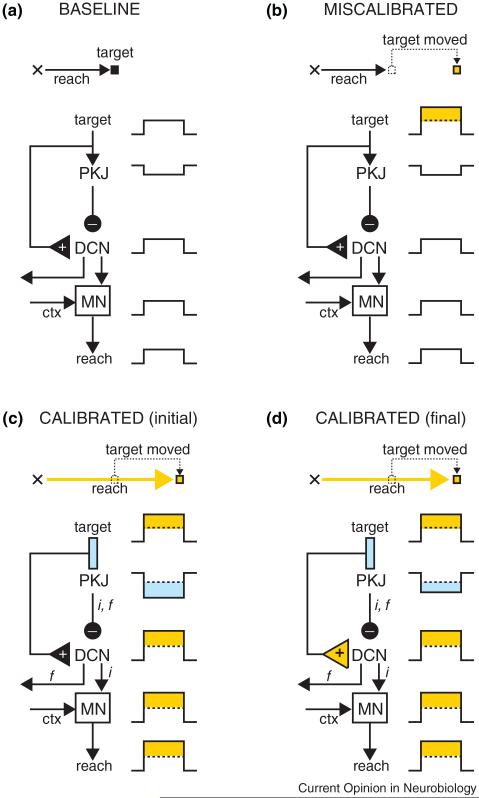
Figure 1.
A hypothesis about sensori-motor calibration after a sensory disturbance. In the baseline phase (a), the hand starts at the position indicated by the cross and the reach takes it to the target location. The activity of Purkinje cells (PKJ), deep cerebellar nucleus (DCN), and motor neurons (MN) is indicated schematically as either increases or decreases in firing rate throughout the entire reach. In (b), the target is shifted to the right (indicated in orange) and as a result the reach falls short. During the early stages of calibration (c), the kinematics of the reach have been altered (indicated in orange) and the hand is moved to the new target location. Note that Purkinje cell activity has changed (cyan) and that it is collaborating with signals from motor cortex (ctx) to improve the motor command generated by the MN (inverse model: ‘i’). Consistent with a forward model (‘f’), the change in Purkinje cell activity is also a prediction of sensory input (i.e. the target will move), and future movement (i.e. the reach will go further). The output of the forward model could be sent out by the DCN to cortex for further processing and state estimation. After extensive training in this task (d), there is a transfer of plasticity from cerebellar cortex to DCN (orange triangle). However in this task, the amount of transfer is small, and Purkinje cell output remains largely unaffected relative to (c).

