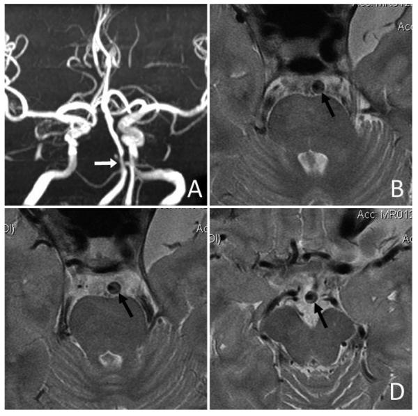
Figure 1.

Intracranial plaque and arterial wall imaging by high-resolution MRI. An ICAS lesion located at proximal basilar artery with severe luminal stenosis was identified on time-of-flight MRA (white arrow in Panel A). High-resolution MRI revealed an eccentric atherosclerotic plaque along the anterolateral and posterolateral walls of basilar artery (black arrows in Panels B, C and D). (Courtesy of Professor WH Xu of Peking Union Medical College Hospital, Beijing, China.)
