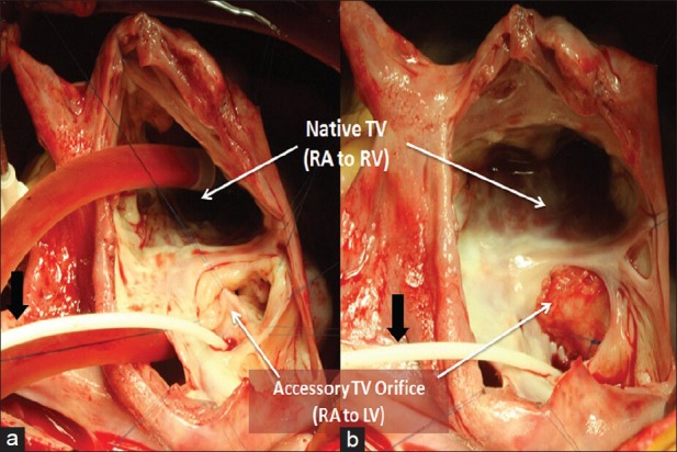
Figure 2.

(a) Surgeon's view through right atriotomy, showing an accessory tricuspid valve (TV) orifice; black arrow pointing to the catheter positioned through the coronary sinus; (b) Surgeon's view through right atrium, after closure of the pericardial patch of the accessory tricuspid orifice; black arrow pointing to the catheter positioned through the coronary sinus
