. 2014 Mar 18;3:e01939. doi: 10.7554/eLife.01939

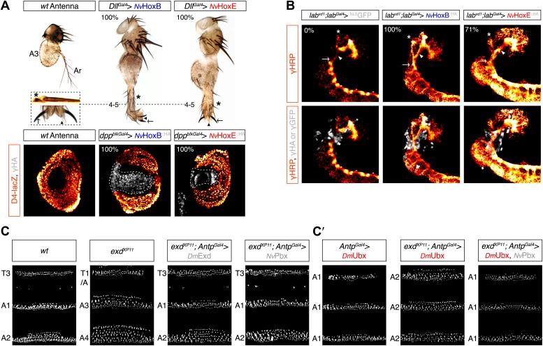
Figure 6. Functional analysis of NvHox and NvPbx proteins in Drosophila.

(A) Antenna-to-leg transforming activities of NvHoxB and NvHoxE. NvHox proteins were expressed in the antenna with the Distalless (Dll)-Gal4 driver. Asterisk depicts leg-specific bracted bristles. 4–5 shows the transformation of the arista in two tarsal segments. Arrow and arrowhead in the enlargement indicate the formation of the leg-specific terminal claw and its associated sensory pad respectively. The antenna-to-leg transformation by NvHox proteins (grey) is achieved through the repression of the spineless (ss) target gene, as observed by the repression of the ss enhancer D4 activity on lacZ reporter gene expression (orange). See also Figure 6—figure supplements 1 and 2. (B) Rescue of the labial (lab) mutant phenotype in the tritocerebrum by NvHox proteins. The central nervous system is stained with an anti-HRP (orange). Hox or GFP (as a control) proteins (grey) are expressed in the tritocerebrum with a lab-Gal4 driver. Frontal connectives (asterisk), longitudinal connectives (arrowhead) and tritocerebral commissure (arrow) are indicated. In lab mutant background, longitudinal connectives are reduced, frontal connectives project ectopically and the tritocerebral commissure is missing (Hirth et al., 1998). Expression of NvHoxB or NvHoxE in this mutant context leads to a complete or strong rescue of this phenotype, respectively. See also Figure 6—figure supplement 2. (CC′) NvPbx can rescue zygotic exd mutant phenotypes in the Drosophila larva cuticle. (C) Larvae homozygous for the zygotic exdXP11 mutation have T3 and A1 segments that resemble to a T1/abdominal or A3 segment, respectively. Thoracic expression of either DmExd or NvPbx in this mutant background (through the UAS/Gal4 system, with the Antennapedia (Antp)-Gal4 driver) is sufficient to restore the correct specification of T3 and A1, as assessed by the shape and arrangement of denticle belts. (C′) Ubx normally specifies the A1 segment. Ectopic expression of Ubx with Antp-Gal4 induces A1-like segments in the thorax. In absence of Exd, Ubx produces A2-like segments. Providing back NvPbx in this genetic background is sufficient to restore the normal A1-inducing activity of Ubx. See also Figure 6—figure supplements 3 and 4.

DOI: http://dx.doi.org/10.7554/eLife.01939.015

Figure 6.

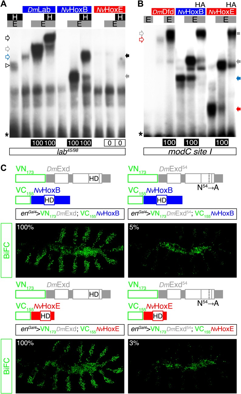
Figure 6—figure supplement 1. NvHoxB and NvHoxE interact with the Drosophila TALE cofactors Extradenticle (Exd) and Homothorax (Hth) in vitro and in vivo.

Figure 6—figure supplement 1.

(A and B) Band shift experiments on physiological target sequences of Drosophila Hox proteins. Probes derived from the auto-regulatory element of the Hox genes labial (lab48/95, A) and Deformed (Dfd, modCsite 1, B), corresponding to binding sites of class I or II, respectively. Colour codes and annotations are as in Figure 5—figure supplement 2. Unfilled arrows indicate DNA-binding complexes with Drosophila Hox (DmLab and DmDfd) and TALE (E and H) proteins. Grey scare in (B) indicates the supershift band resulting from the addition of an antibody against the HA tag of NvHox proteins. (C) Visualisation of interactions between NvHox proteins and Exd by BiFC in a live stage 10 Drosophila embryo. Fusion constructs are schematized above each picture. Constructs were expressed with the engrailed (en)-Gal4 driver. Specificity of BiFC was verified with the Exd fusion construct mutated in the residue 54 of the HD.
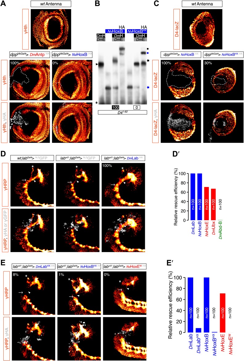
Figure 6—figure supplement 2. Role of the HX of NvHox proteins in generic Drosophila Hox assays.

Figure 6—figure supplement 2.

(A) Antenna-to-leg transforming activities of NvHox proteins do not rely on the repression of homothorax (hth) target gene. Expression of NvHoxB (grey) in the proximal part of the antenna imaginal disc has no effect on hth expression (as assessed by an immunostaining against Hth, orange). By comparison, expressing the Drosophila Antennapedia (Antp) in the same condition leads to a complete loss of Hth. (B) The HX mutation abolishes complex formation between NvHoxB and Drosophila TALE cofactors on the target binding sites of the spineless D4 regulatory element. Colour codes and annotations are as in Figure 5—figure supplement 2. (C) The HX mutation affects the repressive activity of NvHoxB on the spineless D4 target element in vivo. Proteins were expressed as in (A). Expression of D4 was indirectly quantified through immunostaining against the D4-driven LacZ reporter protein (orange). (DE′) The HX mutation abolishes the rescue activity of Drosophila and Nematostella Hox proteins in the tritocerebrum of labial (lab) mutant embryos. (DD′) Rescue assays with wild-type Hox proteins, as indicated. (EE′) Rescue assays with HX-mutated Hox proteins, as indicated. The central nervous system is stained with an anti-HRP (orange). HA-tagged proteins (grey) were expressed in the tritocerebrum with a lab-Gal4 driver. For quantifications of rescue efficiency (D′E′), 100 embryos were examined in each case. Percentage values were corrected in order to take into account the phenotypic penetrance of the lab mutation in tritocerebral development (90%). The rescue efficiency of Dmlab was used as a standard and taken as 100%. The rescue values (the relative percentage of embryos showing a complete rescue of the tritocerebral brain defects) of DmHox and NvHox gene products are shown in percentage relative to Dmlab.
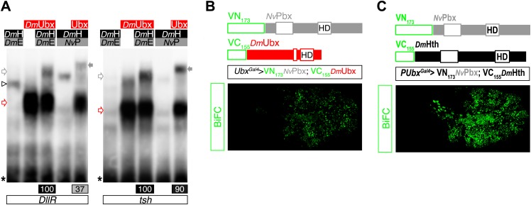
Figure 6—figure supplement 3. NvPbx interacts with the Drosophila Ultrabithorax (Ubx) and Homothorax (Hth) proteins in vitro and in vivo.

Figure 6—figure supplement 3.

(A) Band shift experiments on DNA-binding sites derived from the regulatory elements of the Distalless (DllR) and teashirt (tsh) Hox target genes. Colour code and annotations are as in previous figures. (BC) NvPbx interacts with Ubx (B) and Hth (C) in vivo. Fusion proteins generated for BiFC are schematized above the pictures. Expression was conducted with the Ubx-Gal4 driver. Pictures were acquired in a live stage 10 embryo.
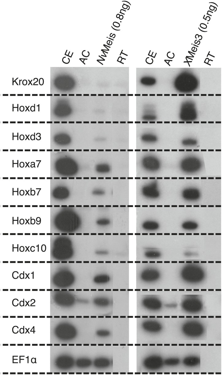
Figure 6—figure supplement 4. NvMeis reproduces generic bilaterian Meis activities in the Xenopus embryo.

Figure 6—figure supplement 4.

While more anterior hindbrain marker genes are not expressed (krox20, hoxd1 and hoxd3), more posterior spinal cord marker genes such as, hoxa7, hoxb7, hoxb9, hoxc10, hoxd10, cdx1, cdx2 and cdx4 are robustly induced upon injection of NvMeis in the embryo. Total RNA was isolated from five control embryos (CE) and eighteen animal caps (AC) from the control or injected groups. Semi-quantitative RT-PCR analysis was performed on AC explants that were removed from control (non injected, RT) and injected embryos cultured until the neurula stage 16. EF1αcontrols for RNA levels in each sample. A comparative level of genes induction by XMeis3 is provided on the right.