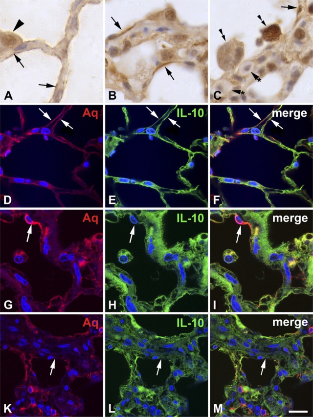
Figure 2.

IL-10 is expressed in type I pneumocytes of the normal rat lung (A, D—F) and in the rat lung with radiation-induced fibrosing alveolitis 3 months after irradiation (B, C, G—M). Rat lungs were formalin-fixed and subjected to immunohistochemistry and immunofluorescence. IL-10 is expressed on the surface of the alveolar walls (arrows in A, E, F) of the normal lung, suggesting positivity of type I pneumocytes. Type II pneumocytes (arrowhead) are also positive, both in the cytoplasm and in the nucleus. In the lung with fibrosing alveolitis, the alveolar walls are thickened and the surface contains IL-10—positive cells that are thicker and do not cover the whole surface of the alveoli (arrows in B, C). Alveolar macrophages (double arrows in C) adhere to areas of the alveolar surface that lack IL-10. Mast cells (arrowheads with asterisk) contain low amounts of IL-10. Aquaporin-5 (Aq), a marker of type I pneumocytes, is colocalized with IL-10 in normal lung (arrows in D—F). In the lung with fibrosing alveolitis (G—M), partially differentiated type I pneumocytes cover a smaller surface than in the control lung, but they have a greater thickness and contain less IL-10 (arrows in H, I). There are areas of the alveolar surface that contain no type I pneumocytes (arrows in K—M). Bars: A = 6.4 μm; B, C = 12.9 μm; D—F = 16.75 μm; G—I = 16.9 μm; K—M = 25.8 μm.
