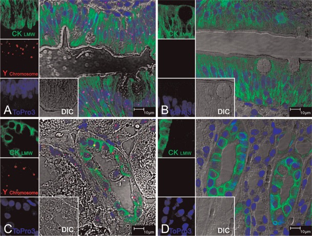
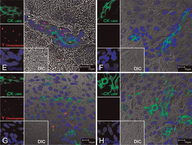
Figure 1.
Dual Y chromosome fluorescence in situ hybridization (FISH) with cytokeratin 8/18 (CK8/18) immunofluorescence staining on normal rhesus monkey tissues. Multilabel confocal microscopy in normal male rhesus monkey tissues: (A) jejunum, (C) kidney, (E) liver, and (G) pancreas; and female rhesus monkey tissues: (B) jejunum, (D) kidney, (F) liver, and (H) pancreas. Images for individual channels [epithelium marker CK8/18 with Alexa 488 is green, Y chromosome FISH with Fast Red is red, nucleus marker To-pro-3 is blue, and differential interference contrast (DIC) is gray] are shown on the left and bottom, and a large merged image containing three channels plus DIC showing double-positive cells is shown on the right. The pink/fuchsia spots (due to the combination of red and blue) indicate Y chromosome signals, which are located inside the blue-staining nucleus, as shown in A, C, E, and G. There were no Y chromosome paintings in the corresponding female tissues used as controls, as shown in B, D, F, and H. Immunofluorescence staining with CK8/18 was shown in all tissues used: the jejunum intestinal epithelium (A, B); the distal tubule epithelium in kidney (C, D); the bile duct epithelium of liver (E, F); and the intralobular duct epithelium in pancreas (G, H). The DIC images indicated that the morphology of all tissues was well preserved.


