
Figure 2.
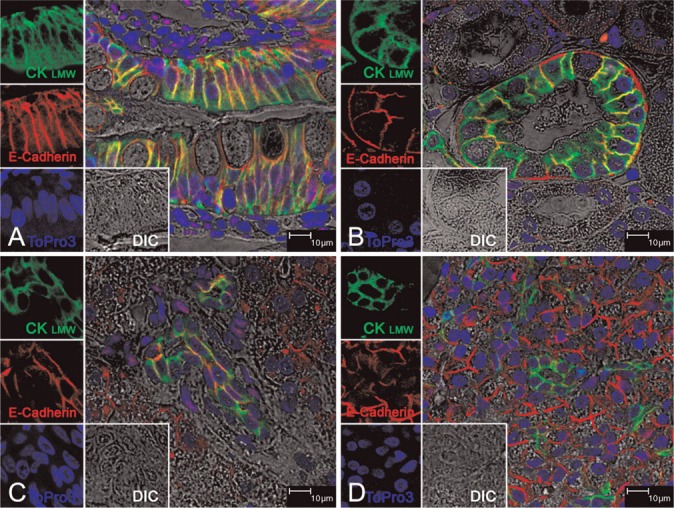
Immunofluorescence staining of CK8/18 on healthy rhesus monkey tissues. All panels show triple-labeled confocal microscopy (four channels). Images for individual channels [CK8/18 with Alexa 488 is green, E-cadherin (E-cad) with Alexa 568 is red, To-pro-3 is blue, and DIC is gray] are shown on the left and the main panel shows the merged image containing all three channels plus DIC. The areas of green and red fluorescence overlap are seen as yellow. (A) Jejunum epithelial cells; (B) kidney distal tubule epithelial cells; (C) bile duct epithelial cells of liver; (D) intralobular ductal epithelial cells of pancreas. Structures are all predominantly stained for CK8/18 and E-cad, whereas E-cad red fluorescence but not CK8/18 is seen on hepatocytes in C and acinar cells of pancreas in D. Taken together, CK8/18 is seen exclusively to stain epithelial cell structures of all of these tissues.