
Figure 1.
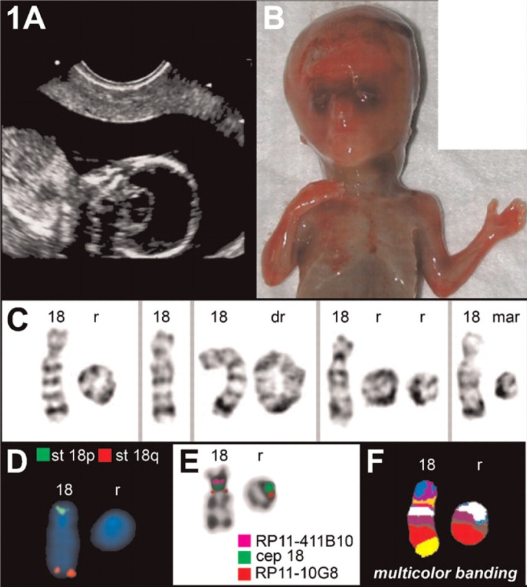
Case A. (A) Sonography revealed a holoprosencephaly (HPE). (B) Frontal view of the autopsy of the fetus at 17 weeks, showing the HPE. (C) Partial G-banded karyotype showing the normal chromosome 18, the single ring (r), double ring (dr), two rings, and the marker (mar) (D) Commercially available FISH subtelomeric probes (Abbott/Vysis) for chromosomes 18p (st 18p) and 18q (st 18q) demonstrated that both corresponding regions are deleted in the ring chromosome. (E) Fluorescence in situ hybridization applying subcentromeric probes revealed that the centromere-near region in 18p11.21 was absent and the centromere-near region in 18q11.21 was present on the ring. (F) Multicolor banding (MCB) confirmed the aforementioned findings.