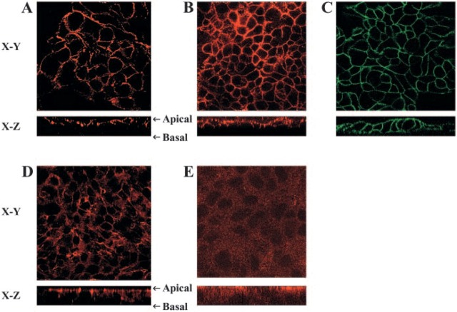
Figure 2.
Surface distribution of invariant chain (Ii) on N87 cells grown on filter inserts. Confocal microscopy images are shown here of the horizontal interfaces (X-Y) and vertical interfaces (X-Z). (A) N87 cells stained with anti-DAF antibody (Alexa 568) as an apical marker, (B) N87 cells stained with anti-Ii antibody (Alexa 568), and (C) N87 cells stained with anti-epithelial antigen with apical and basal staining (FITC). Cells were stained with saturating concentration of antibodies, which reach both apical and baso-lateral sides of the cells. As additional controls, the cells were stained with antibodies to (D) the apical junctional complex protein Zona Occludens-1 and a secondary goat anti-mouse IgG conjugated with Alexa 568 and to (E) the cytoplasmic tail of Ii also with the same secondary antibody conjugated with Alexa 568.

